Ultrasound
Ultrasound is an imaging technique that uses sound waves to view soft-tissue organs within the body.
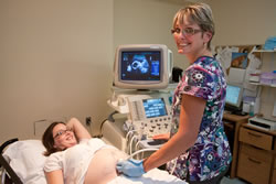 An ultrasound study begins by applying a water-soluble gel to the appropriate part of the body, enabling transmission of sound waves from a transducer (or camera) as it moves across the patient's skin.
The technologist who interprets and conducts this test is called a sonographer and the physician who reports the study is called a radiologist. All of the sonographers who work at RVH have completed qualifying exams and are registered with the College of Medical Radiation and Imaging Technologists of Ontario. Our expert staff specialists, employing appropriate ultrasound equipment and techniques, ensure that we provide you with the best diagnostic assessment to support you and your doctor.
There are no known side effects from ultrasound and this is why it is an excellent tool to image the fetus. Obstetrical ultrasound makes up a large portion of our work, in addition to investigations of the abdomen and other soft tissue organs. Ultrasound can also be used to assist with various types of biopsies, as well as imaging blood vessels and analyzing blood flow.
Three of the most common ultrasound procedures are abdominal and pelvic ultrasound, obstetrical and pelvic ultrasound and renal ultrasound (kidney and bladder). RVH also provides Echocardiography which is an ultrasound of the heart.
Visiting or accompanying patients
You are very welcome to bring one adult friend or family member along to your ultrasound appointment. In the case of an obstetrical ultrasound, which we try to make a warm bonding event, we invite you to include one other person you may wish to share this moment with. They are welcome into the room after the diagnostic portion of the ultrasound is complete. Children (12 and under) should not accompany the patient to any ultrasound appointment, as we cannot provide for their supervision while completing your test or treatment.
What you should know
When you come to the hospital, please arrive 15 minutes early to register at the Medical Imaging Reception Desk. Bring your health card.
Where appropriate, we can offer you a copy of your obstetrical ultrasound image.
Abdominal Ultrasound - FAQ's
If you are diabetic, please let the booking clerk know at the time of booking to ensure an early morning appointment.
What is an abdominal ultrasound?
During an abdominal ultrasound we may assess the anatomical integrity of a number of different soft-tissue organs, including the liver, gallbladder, kidneys, pancreas, spleen and major veins and arteries. An abdominal ultrasound encompasses the area below the lung and to the approximate area of the belly button.
What do I need to do before the ultrasound?
Have nothing to eat or drink for eight hours prior to your appointment (except to swallow necessary medications). For children under three years, feed as usual. Your empty stomach is much less gaseous than it is after you've eaten. An empty stomach results in a much more clear image for the radiologist. An empty stomach also helps us adequately visualize the gall bladder, which contracts when a person eats.
Obstetrical and pelvic Ultrasound - FAQ's
What is an Obstetrical and Pelvic Ultrasound?
During an Obstetrical and Pelvic Ultrasound we may include the following in our assessment: gestational age, placental position, appropriate growth indices, fetal anatomy and fetal well being.
What do I need to do before the ultrasound?
For obstetrical examinations before 12 weeks, or for pelvic ultrasound, a full bladder is necessary. The pelvic organs are situated behind the urinary bladder, and when full it acts as a window for the sound waves. Please finish drinking four full 8 oz glasses of clear fluid (not milk or tomato juice) at least one hour prior to your appointment. Do not empty your bladder after drinking the fluids until completion of the exam - a washroom is close by the examining room!
For obstetrical examinations later than 12 weeks, a full bladder is not necessary. No special preparation is required. Some examinations include a transvaginal ultrasound, where we insert a thin probe into the vagina to allow better visualization of the anatomy. Patients are able to void prior to this procedure.
Renal Ultrasound (Kidney and Bladder) - FAQ's
What do I need to do before the ultrasound?
Drink two full 8 oz. glasses of clear fluid to fill your bladder. Please do not void until after the examination.